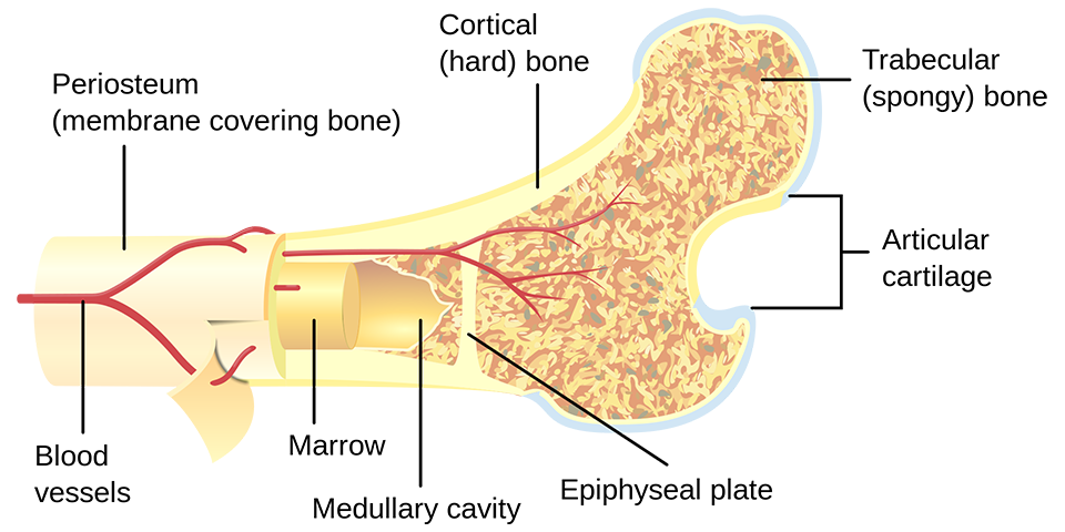
Cortical Bone
The hard outer layer of bones is composed of cortical bone also called compact bone being much denser than cancellous bone. It forms the hard exterior (cortex) of bones.
The cortical bone gives bone its smooth, white, and solid appearance, and accounts for 80% of the total bone mass of an adult human skeleton. It facilitates
bone's main functions: to support the whole body, protect organs, provide levers for movement, and store and release chemical elements, mainly calcium. It consists of
multiple microscopic columns, each called an osteon. Each column is multiple layers of osteoblasts and osteocytes around a central canal called the haversian canal.
Volkmann's canals at right angles connect the osteons together. The columns are metabolically active, and as bone is reabsorbed and created the nature and location of the
cells within the osteon will change. Cortical bone is covered by a periosteum on its outer surface, and an endosteum on its inner surface. The endosteum is the boundary
between the cortical bone and the cancellous bone. The primary anatomical and functional unit of cortical bone is the osteon.
Cancellous Bone
Cancellous bone, also called trabecular or spongy bone, is the internal tissue of the skeletal bone and is an open cell porous network.
Cancellous bone has a higher surface-area-to-volume ratio than cortical bone because it is less dense. This makes it weaker and more flexible. The greater surface
area also makes it suitable for metabolic activities such as the exchange of calcium ions. Cancellous bone is typically found at the ends of long bones, near joints
and in the interior of vertebrae. Cancellous bone is highly vascular and often contains red bone marrow where hematopoiesis, the production of blood cells, occurs.
The primary anatomical and functional unit of cancellous bone is the trabecula. The trabeculae are aligned towards the mechanical load distribution that a bone
experiences within long bones such as the femur. As far as short bones are concerned, trabecular alignment has been studied in the vertebral pedicle. Thin
formations of osteoblasts covered in endosteum create an irregular network of spaces, known as trabeculae. Within these spaces are bone marrow and hematopoietic
stem cells that give rise to platelets, red blood cells and white blood cells. Trabecular marrow is composed of a network of rod- and plate-like elements that make the overall organ
lighter and allow room for blood vessels and marrow. Trabecular bone accounts for the remaining 20% of total bone mass but has nearly ten times the surface area
of compact bone. The words cancellous and trabecular refer to the tiny lattice-shaped units (trabeculae) that form the tissue. It was first illustrated accurately in the
engravings of Crisóstomo Martinez.